
Discussion
To the best of our knowledge, this is the first nationwide surveillance to examine the prevalence of PAI in patients with type 1 diabetes and clarify the clinical characteristics in these patients. In the present study, we clearly demonstrated that slowly progressive type 1 diabetes and female sex are independently associated with PAI, achieving the ORs of 5.7 and 13.87, respectively, in patients with type 1 diabetes.
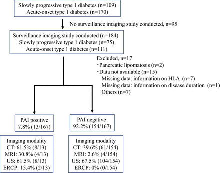
In the present study, pancreatic cystic lesions were identified in 7.7% (5/65) of patients with slowly progressive type 1 diabetes (IPMN, n=1; pancreatic cysts, n=4), a higher prevalence compared with 1.9% (2/102) of patients with acute-onset type 1 diabetes (suspected IPMA, n=1; pancreatic cyst, n=1). IPMN and PanIN are mucinous cystic lesions arising from pancreatic ducts and each well-recognized PADC precursor.18 Our pancreatic exocrine histology has shown that a high proportion of slowly progressive type 1 diabetes pancreas (diabetes duration, 13±7 years) had pancreatic cystic lesions associated with PanIN in the branches and smaller ducts,19 whereas acute-onset type 1 diabetes showed mild periductal fibrosis with less pancreatic ductal morphological changes.20 Additionally, the number of PanIN-positive lobes in the exocrine pancreas increased with increased duration of slowly progressive type 1 diabetes,4 21 which suggests a progressive increase of pancreatic cystic lesions associated with PanIN along with longer duration of diabetes. These results indicate that slowly progressive type 1 diabetes may be more likely to be associated with mucinous cystic precursor lesions relevant to PDAC than acute-onset type 1 diabetes. Recently, we reported that EV encoded-capsid protein 1 was detected in the exocrine as well as endocrine glands of patients with slowly progressive type 1 diabetes with varying duration of diabetes, and the persistent EV infection triggers exocrine pancreatic inflammation and transforms acinar cells into acute-onset type 1 diabetes and PanIN, followed by pancreatic cystic lesions.4 Therefore, a chronic inflammatory condition of the pancreas caused by EV infection may play some roles in the development of pancreatic cystic lesions in slowly progressive type 1 diabetes. Previous research has shown that obesity and/or insulin resistance are correlated with the development of pancreatic cysts in patients with type 2 diabetes,22 yet the mechanistic basis for these relationships is not well understood. However, all patients with pancreatic cysts were less obese. Therefore, the underlying mechanisms responsible for the development of pancreatic cysts might be different between slowly progressive type 1 diabetes and type 2 diabetes. Since our results are derived from a dataset including subjects with a relatively short disease duration, the prevalence of pancreatic cystic lesions, namely PAI, might increase over time in patients with slowly progressive type 1 diabetes.
In this study, important clinical characterization of coexistence of PAI indicating precancerous potential was almost observed in females. To date, the association between type 1 diabetes and pancreatic cancer is not well described. A meta-analysis by Carstensen et al23 showed that the HRs of pancreatic cancer were 1.53 (95% CI 1.30 to 1.79) in males and 1.25 (95% CI 1.02 to 1.53) in females, respectively, indicating non-sex-specific cancers, in patients with type 1 diabetes. Interestingly, it has been reported that the majority of type 1 diabetes subjects with PADC were female by case study in a Japanese population.24 However, the pathophysiological mechanism underlying the higher incidence of PADC—dominant in females—and pronounced gender difference in the frequency of PAI observed in the present study remains unsolved. Therefore, further studies are needed to assess the exact incident rate of PAI including PADC in patients with type 1 diabetes, especially focusing on cases of slowly progressive type 1 diabetes.
Current evidence strongly supports the view that the presence of one or more islet autoantibodies reflects an autoimmune process that precedes and contributes to the development of type 1 diabetes, rather than being a secondary phenomenon resulting from pancreatic damage.25 26 As shown in table 1, there was no significant difference in the positivity rate of islet autoantibodies between the PAI-positive and PAI-negative groups. These findings suggest that the presence of islet autoantibodies is associated with islet-specific autoimmune responses, while PAI may occur independently of such autoimmunity. Further investigations are needed to determine whether the coexistence of PAI affects the islet autoimmunity.
Lastly, the comparisons of combinations of characteristics revealed that female sex and slowly progressive type 1 diabetes are associated with PAI, indicating precancerous potential in patients with type 1 diabetes. Furthermore, pancreatic imaging should be performed at the time of diagnosis in female subjects with slowly progressive type 1 diabetes, and those with PAI should undergo closer monitoring to facilitate early detection of PADC throughout the disease process.
There were several limitations that should be addressed. First, the number of patients was relatively small. Therefore, additional studies with larger numbers of participants with type 1 diabetes will be needed to confirm our results. Second, since conventional imaging modalities of the abdomen, such as US, CT, or MRI, were used as a screening test in this study, the prevalence of PAI may have been underestimated due to the low sensitivity for identifying pancreatic cystic lesions compared with endoscopic US and/or ERCP. The choice of imaging modality was determined by the diabetes specialist at each facility, and no standardized criteria were used. Although imaging was performed in all cases, the variety of modalities used may also represent a limitation. Third, several population-based imaging studies have reported that pancreatic findings, such as cystic lesions and glandular atrophy, increase with age. Some studies have also shown a slightly higher prevalence of these findings in women, even among the general population or individuals without known pancreatic disease.27 28 These observations may be related to physiological ageing processes or sex-related anatomical or hormonal factors. Unfortunately, our study did not include a non-diabetic control group with imaging data for direct comparison. Therefore, although PAI findings were more frequent in elderly individuals and women with type 1 diabetes in the current study, these results may partially reflect general population trends, rather than being specific to type 1 diabetes. Further studies are needed to clarify the role of ageing and sex differences in PAI development in patients with type 1 diabetes.
In conclusion, concomitant PAI may be a distinct clinical feature of slowly progressive type 1 diabetes, compared with acute-onset type 1 diabetes.
Leave a Reply